
1.4 Cerebral Cortex
Section Learning Objectives
1.7 Identify the four lobes of the cortex.
1.8 Describe the behavioral functions associated with each of the four cortical lobes.
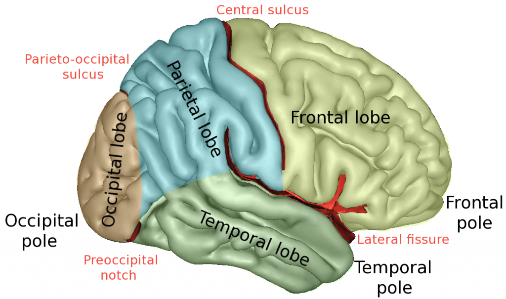
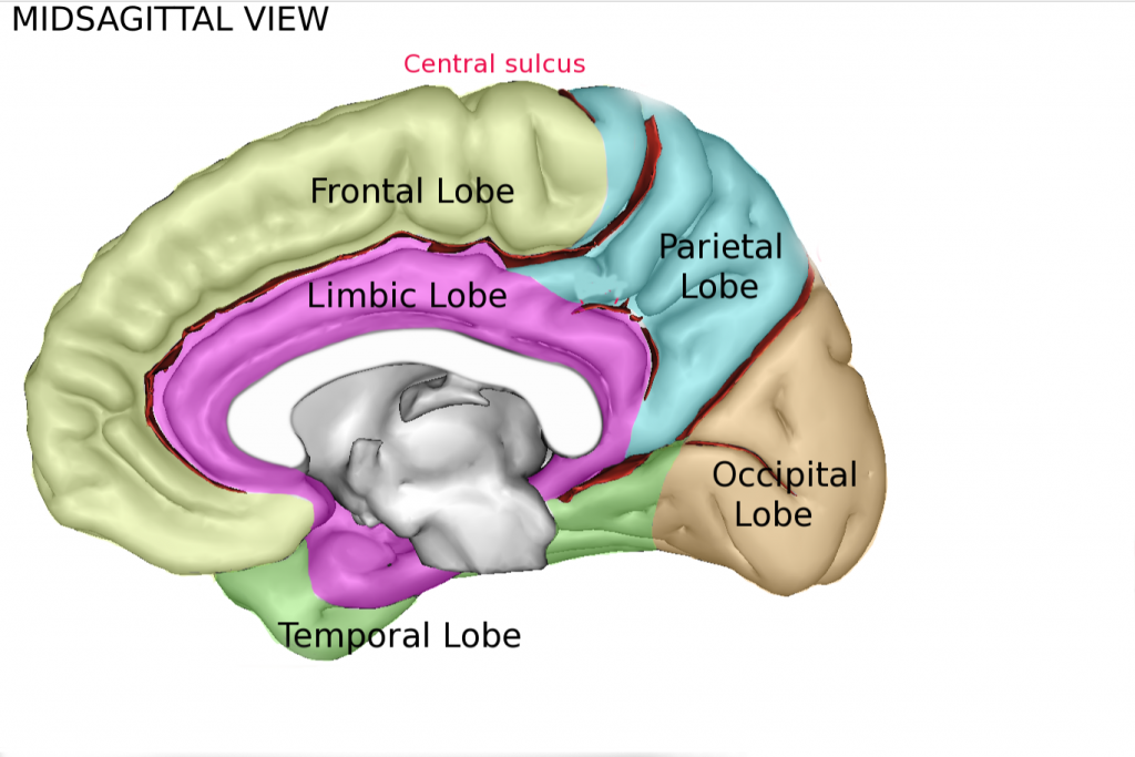
The cerebral cortex is divided into two hemispheres each of which has four lobes: Occipital, Temporal, Parietal, Frontal. These are pictured in the Figure 1.3 and Figure 1.4. The structures of the brain. Each lobe is specialized for processing certain functions. As you learn each of these structures and associated functions, it’s important to keep in mind that the brain is highly interconnected, and signals are constantly sent across the entire brain.
The occipital lobes are located at the posterior (back) of the brain. They are important in processing visual information. The temporal lobes are on the inferior (bottom) of the brain. They play key roles in processing auditory information. The left temporal lobe is specialized for understanding the meanings of language. The parietal lobes are located on the superior (top) of the brain. They receive and process information from our somatosensory system (skin senses) about pain, touch, and temperature. They also integrate information across the senses. For example, hearing a dog barking and seeing it running toward you with its tail wagging. The frontal lobe contains the motor strip that issues action signals to your muscles via the spinal cord. The frontal lobes also control the executive functions that allow for impulse control, abstract thinking, planning and judgement. The left frontal lobe has a specialized area, Broca’s area, that is important in production of speech.
The corpus callosum is a thick band of nerve fibers that connect the two hemispheres. It is important in communication between the two hemispheres.
Exercises
Practice labeling the lobes of the cortex.
agittal perspective. The temporal lobe is shown on the bottom of the image, bordering the occipital and frontal lobes. On the image, the temporal lobe borders the frontal lobe on the left, the frontal lobe extends up and around to the top of the brain in the image. Continuing to the right of the image from the frontal lobe, is the central sulcus border into the parietal lobe region. The parietal lobe then rounds to the right, and down to the occipital lobe. the occipital lobe then connects back to the temporal lobe on the bottom of the image. The temporal lobe is appears separated at the middle by another piece that is unlabeled, and goes toward the middle of the image. The limbic lobe is connected to this piece on both sides, and bordering the inner perimeter of the other 4 lobes.
Links For Learning
For interactive 3D views of the brain explore:
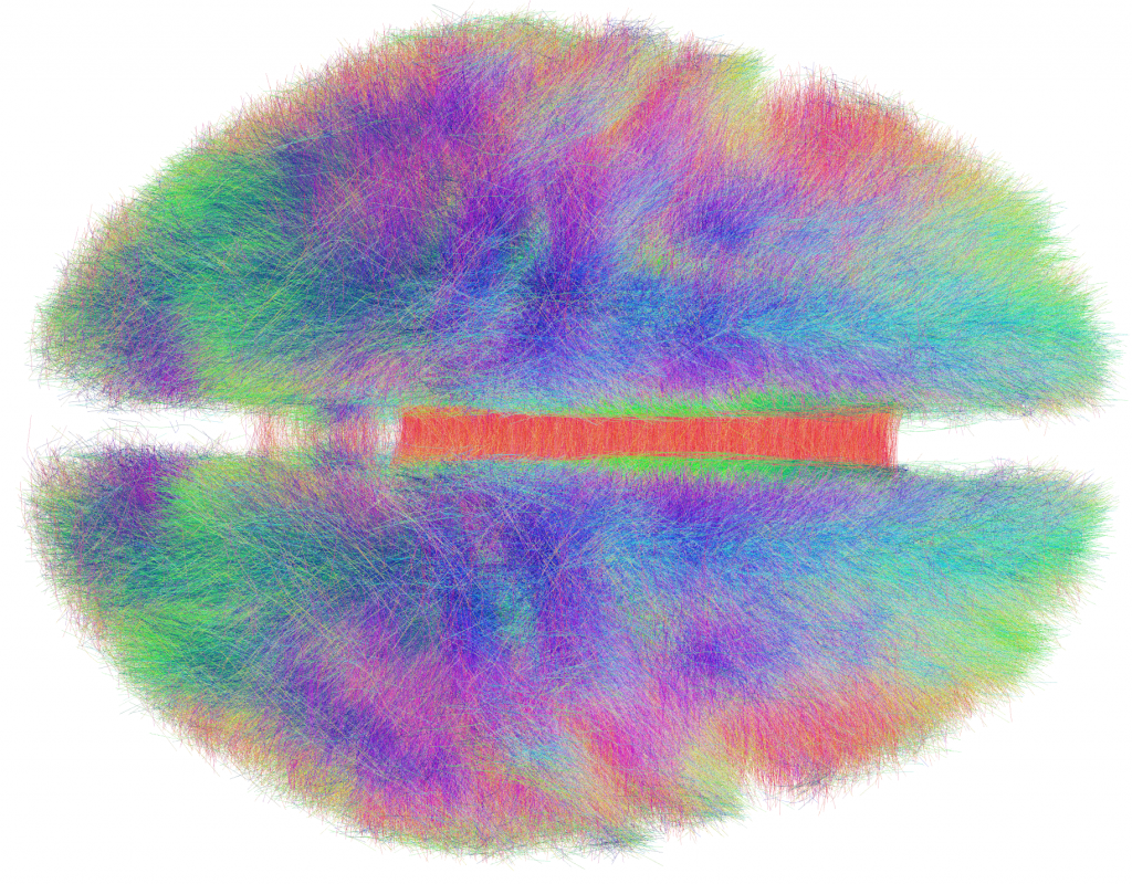
Learning Myth Buster: We only use 10% of our brain.
This myth may have originated from Dale Carnegie’s claim that people only used a part of their brains in an effort to inspire them to reach higher goals. Or it may have come from a misinterpretation of William James statement that people “make use of a very small portion of their possible consciousness and of their soul’s resources, in general.” Jarrett (2014) proposes several possible origins for this myth. These claims, however, are not scientifically sound. As Jarrett points out, that would be inconsistent with evolutionary principles. Current neuroimaging studies show that the brain is highly interconnected. The Human Connectome project is working to map the interconnections of the brain. Figure 1.5 shows one of their images.