
3 3.1 Prenatal Brain Development
Section Learning Objectives
- Describe and compare the processes of migration and differentiation during prenatal brain development
- Identify the progression of the development of the major brain structures through prenatal development.
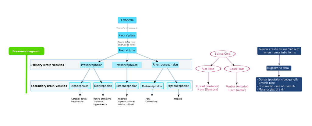
The human brain changes dramatically throughout the course of prenatal development. At its earliest stages, about twenty-five days after conception, it is but a thin sheet of cells. This sheet rolls upon itself to become the neural tube. This tube differentiates to form the three major divisions of the brain (Prosencephalon, mesencephalon, and rhombencephalon). These divisions are noticeable by 40 days of gestation, but the proportions are much more equal than that of a developed brain. These three subdivisions further divide and specialize. This is summarized in Figure 3.1. The prosencephalon subdivides to the telencephalon, which becomes the forebrain, and diencephalon (which develops into the thalamas and hypothalamus). The mesencephalon develops to become the midbrain. The rhombencephalon, or hindbrain, forms the brainstem including the medulla, pons, and cerebellum. The gyri and sulci begin to form by the seventh month. The brain at birth has all the major components of the adult brain, but does not yet function the same.
Long Description
A flowchart describing the development of the nervous system
Title: Development of Nervous System
At the left is Foramen magnum. To the right are 3 flowcharts.
The first flowchart starts with Ectoderm. Ectoderm thickens to become the Neural plate. Neural folds rise and fuse to form the Neural tube.
The Neural tube points to 3 parts of the Primary Brain Vesicles: the Prosencephalon, the Mesencephalon and the Rhombencephalon.
This flowchart continues from the Primary to the Secondary Brain Vesicles.
The Prosencephalon points to 2 parts of the Secondary Brain Vesicles: the Telencephalon and the Diencephalon. The Mesencephalon points to the Mesencephalon in the Secondary Brain Vesicles. The Rhombencephalon points to the Metencephalon and Myelencephalon in the Secondary Brain Vesicle.
From the Secondary Brain Vesicles, the Telencephalon points to the cerebral cortex basal nuclei. The Diencephalon points to the retina of the eye, the thalamus and hypothalamus. The Mesencephalon points to the Midbrain, the superior colliculi and the inferior colliculi. The Metencephalon points to the pons and cerebellum. The Myelencephalon flows to the Medulla.
The second flowchart begins with the Spinal Cord which points to 2 areas: the Alar Plate and the Basal Plate. The Alar Plate then points to the Dorsal (posterior) and Horn [Sensory], while the Basal Plate points to the Ventral (Anterior) and Horn [Motor].
At the far right is the last flowchart.
“Neural crest is tissue ’left out” when neural tube forms.” This points to
“Migrates to form” which points to
- Dorsal (posterior) root ganglia
- Enteric plexi
- Chromaffin cells of medulla
- Melanocytes of skin
During prenatal development, the brain follows very specific patterns. New cells are formed through the process of neurogenesis and gliogenesis. As the nerve and glial cells are formed, their roles are predetermined. As the cells are formed, they migrate to their predetermined location in the brain. This is called the process of cell migration. The cells then take on specific roles through the process of cell differentiation. Neural maturation occurs after the cells have migrated to their ultimate position within the brain. The maturation process includes making connections to other neurons through the formation of synapses. Dendrites branch out and dendritic spines form. This process begins prenatally, but continues after birth. Dendrites grow more slowly than axons. Growth of the dendrites and axons can be influenced by both genetic and environmental factors. These environmental factors include the prenatal environment.
Link to Learning