
Eukaryotes
8.4 – Ecology of Protists
Learning Objectives
By the end of this section, you will be able to do the following:
- Describe the role that protists play in the ecosystem
- Describe important pathogenic species of protists
Protists function in various ecological niches. Whereas some protist species are essential components of the food chain and generators of biomass, others function in the decomposition of organic materials. Still other protists are dangerous human pathogens or causative agents of devastating plant diseases.
Primary Producers/Food Sources
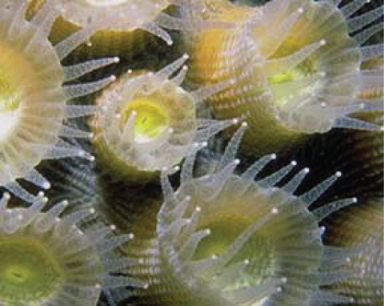
Protists are essential sources of food and provide nutrition for many other organisms. In some cases, as with zooplankton, protists are consumed directly. Alternatively, photosynthetic protists serve as producers of nutrition for other organisms. Paramecium bursaria and several other species of ciliates are mixotrophic due to a symbiotic relationship with green algae. This is a temporary version of the secondarily endosymbiotic chloroplast found in Euglena. But these symbiotic associations are not restricted to protists. For instance, photosynthetic dinoflagellates called zooxanthellae provide nutrients for the coral polyps ((Figure)) that house them, giving corals a boost of energy to secrete their calcium carbonate skeleton. In turn, the corals provide the protist with a protected environment and the compounds needed for photosynthesis. This type of symbiotic relationship is important in nutrient-poor environments. Without dinoflagellate symbionts, corals lose algal pigments in a process called coral bleaching, and they eventually die. This explains why reef-building corals typically do not reside in waters deeper than 20 meters: insufficient light reaches those depths for dinoflagellates to photosynthesize.
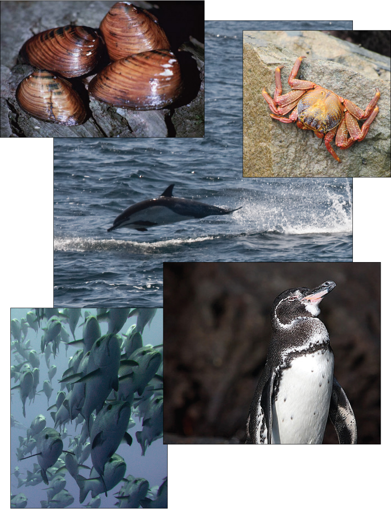
The protists and their products of photosynthesis are essential—directly or indirectly—to the survival of organisms ranging from bacteria to mammals ((Figure)). As primary producers, protists feed a large proportion of the world’s aquatic species. (On land, terrestrial plants serve as primary producers.) In fact, approximately 25 percent of the world’s photosynthesis is conducted by photosynthetic protists, particularly dinoflagellates, diatoms, and multicellular algae.
Protists do not create food sources only for sea-dwelling organisms. Recall that certain anaerobic parabasalid species exist in the digestive tracts of termites and wood-eating cockroaches, where they contribute an essential step in the digestion of cellulose ingested by these insects as they consume wood.
Human Pathogens
As we have seen, a pathogen is anything that causes disease. Parasitic organisms live in or on a host organism and harm the organism. A small number of protists are serious pathogenic parasites that must infect other organisms to survive and propagate. For example, protist parasites include the causative agents of malaria, African sleeping sickness, amoebic encephalitis, and waterborne gastroenteritis in humans. Other protist pathogens prey on plants, effecting massive destruction of food crops.
Plasmodium Species
In 2015 WHO reported over 200 million cases of malaria, mostly in Africa, South America, and southern Asia. However, it is not well known that malaria was also a prevalent and debilitating disease of the North Central region of the United States, particularly Michigan, with its thousands of lakes and numerous swamps. Prior to the civil war, and the drainage of many swamps, virtually everyone who immigrated to Michigan picked up malaria (ague as it was called in the late 1800s), and the pale, sallow, bloated faces of that period were the rule. The only healthy faces were worn by those immigrants who had just arrived. In fact, there were more deaths due to malaria in Michigan than those from the Civil War.
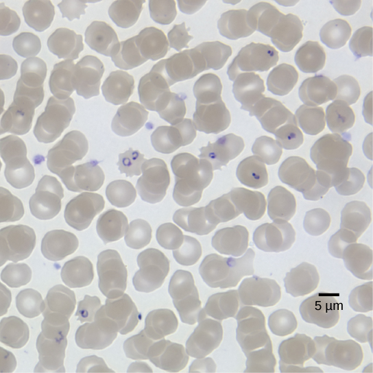
We now know that malaria is caused by several species of the apicomplexan protist genus Plasmodium. Members of Plasmodium must sequentially require both a mosquito and a vertebrate to complete their life cycle. In vertebrates, the parasite develops in liver cells (the exoerythrocytic stage) and goes on to infect red blood cells (the erythrocytic stage), bursting from and destroying the blood cells with each asexual replication cycle ((Figure)). Of the four Plasmodium species known to infect humans, P. falciparum accounts for 50 percent of all malaria cases and is the primary (and deadliest) cause of disease-related fatalities in tropical regions of the world. In 2015, it was estimated that malaria caused over 400,000 deaths, mostly in African children. During the course of malaria, P. falciparum can infect and destroy more than one-half of a human’s circulating blood cells, leading to severe anemia. In response to waste products released as the parasites burst from infected blood cells, the host immune system mounts a massive inflammatory response with episodes of delirium-inducing fever (paroxysms) as parasites lyse red blood cells, spilling parasite waste into the bloodstream. P. falciparum is transmitted to humans by the African mosquito, Anopheles gambiae. Techniques to kill, sterilize, or avoid exposure to this highly aggressive mosquito species are crucial to malaria control. Ironically, a type of genetic control has arisen in parts of the world where malaria is endemic. Possession of one copy of the HbS beta globin allele results in malaria resistance. Unfortunately, this allele also has an unfortunate second effect; when homozygous it causes sickle cell disease.
Link to Learning
This movie depicts the pathogenesis of Plasmodium falciparum, the causative agent of malaria.
Trypanosomes
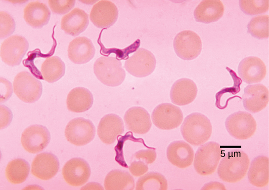
Trypanosoma brucei ((Figure)), transmitted by tsetse flies (Glossina spp) in Africa, and related flies in South America, is an flagellated endoparasite responsible for the deadly disease nagana in cattle and horses, and for African sleeping sickness in humans. This trypanosome confounds the human immune system by changing its thick layer of surface glycoproteins with each infectious cycle. (The glycoproteins are identified by the immune system as foreign antigens, and a specific antibody defense is mounted against the parasite.) However, T. brucei has thousands of possible antigens, and with each subsequent generation, the protist switches to a glycoprotein coating with a different molecular structure. In this way, T. brucei is capable of replicating continuously without the immune system ever succeeding in clearing the parasite. Without treatment, T. brucei attacks red blood cells, causing the patient to lapse into a coma and eventually die. During epidemic periods, mortality from the disease can be high. Greater surveillance and control measures lead to a reduction in reported cases; some of the lowest numbers reported in 50 years (fewer than 10,000 cases in all of sub-Saharan Africa) have happened since 2009.
Link to Learning
This movie discusses the pathogenesis of Trypanosoma brucei, the causative agent of African sleeping sickness.
In Latin America, another species of trypanosome, T. cruzi, is responsible for Chagas disease. T. cruzi infections are mainly caused by a blood-sucking “kissing bug” in the genus Triatoma. These “true bugs” bite the host during the night and then defecate on the wound, transmitting the trypanosome to the victim. The victim scratches the wound, further inoculating the site with trypanosomes at the location of the bite. After about 10 weeks, individuals enter the chronic phase but most never develop further symptoms. In about 30 percent of cases, however, the trypanosome causes further damage, especially to the heart and digestive system tissues in the chronic phase of infection, leading to malnutrition and heart failure due to abnormal heart rhythms. An estimated 10 million people are infected with Chagas disease, and it caused 10,000 deaths in 2008.
Plant Parasites
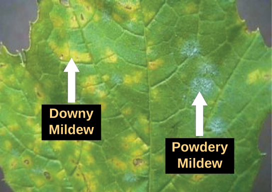
Protist parasites of terrestrial plants include agents that destroy food crops. The oomycete Plasmopara viticola parasitizes grape plants, causing a disease called downy mildew ((Figure)). Grape plants infected with P. viticola appear stunted and have discolored, withered leaves. The spread of downy mildew nearly collapsed the French wine industry in the nineteenth century.
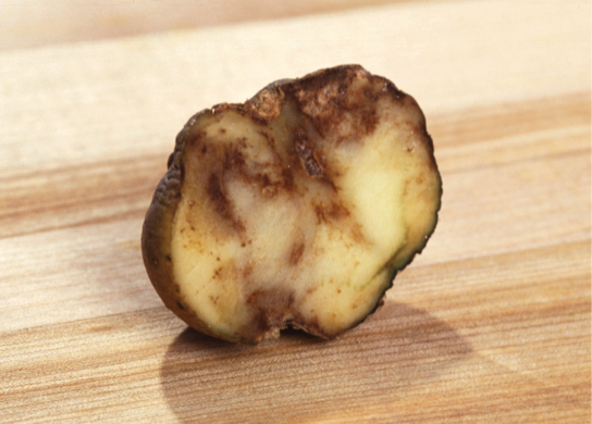
Phytophthora infestans is an oomycete responsible for potato late blight, which causes potato stalks and stems to decay into black slime ((Figure)). Widespread potato blight caused by P. infestans precipitated the well-known Irish potato famine in the nineteenth century that claimed the lives of approximately 1 million people and led to the emigration of at least 1 million more from Ireland. Late blight continues to plague potato crops in certain parts of the United States and Russia, wiping out as much as 70 percent of crops when no pesticides are applied.
Protist Decomposers
The fungus-like protist saprobes are specialized to absorb nutrients from nonliving organic matter, such as dead organisms or their wastes. For instance, many types of oomycetes grow on dead animals or algae. Saprobic protists have the essential function of returning inorganic nutrients to the soil and water. This process allows for new plant growth, which in turn generates sustenance for other organisms along the food chain. Indeed, without saprobe species, such as protists, fungi, and bacteria, life would cease to exist as all organic carbon became “tied up” in dead organisms.
Section Summary
Protists function at several levels of the ecological food web: as primary producers, as direct food sources, and as decomposers. In addition, many protists are parasites of plants and animals and can cause deadly human diseases or destroy valuable crops.
