
Chapter 13: Positive Emotions
Brain Activation – Consistency and Discriminable Brain Patterns
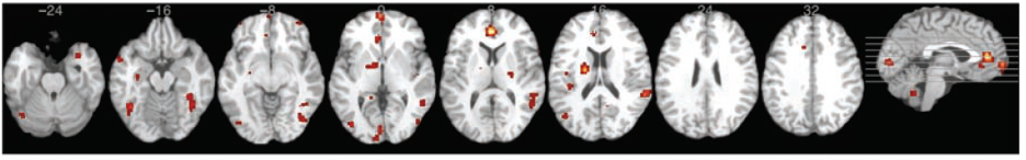
Long Description
The image displays a series of nine horizontal brain scans arranged side by side against a black background. Each scan represents a different cross-sectional view of the brain, progressing from the lower to upper regions. The scans are grayscale, highlighting brain structures, with scattered red and orange markings indicating areas of interest or activity. Numerical labels above each scan (-24, -16, -8, 0, 8, 16, 24, 32) indicate the relative positioning of each slice. On the far right, there is a sagittal view of the brain showing the side profile. The same red and orange markings are visible in this view, highlighting specific regions.
Adapted from “Neuroimaging support for discrete neural correlates of basic emotions: a voxel-based meta-analysis,” by K. Vytal and S. Hamann, 2010, . Journal of Cognitive Neuroscience, 22(12), p. 2870 (https://doi.org/10.1162/jocn.2009.21366). Copyright 2012 by Massachusetts Institute of Technology.
Discriminable Patterns for happiness are shown in Table 24. These findings indicate that compared to other emotions, happiness resulted in more activation in the ACC and STG.
Table 24
Discriminable Patterns for Happiness from Vytal and Hamann (2010)
| Comparison | Happiness Resulted in more Activation in… |
|---|---|
| Happiness vs. Disgust | Left Rostral ACC |
| Happiness vs. Sadness | Right STG (BA 22) |
| Happiness vs. Fear | Right STG (BA 22) |
| Happiness vs. Anger | Left Rostral ACC |
Note: ACC = Anterior Cingulate Cortex, STG= Superior Temporal Gyrus, BA = Brodmann’s Area. Adapted from “Neuroimaging support for discrete neural correlates of basic emotions: a voxel-based meta-analysis,” by K. Vytal and S. Hamann, 2010, Journal of Cognitive Neuroscience, 22(12), p. 2872 – 2874 (https://doi.org/10.1162/jocn.2009.21366). Copyright 2012 by Massachusetts Institute of Technology.
