
Chapter 7: Physiological Measures of Emotion
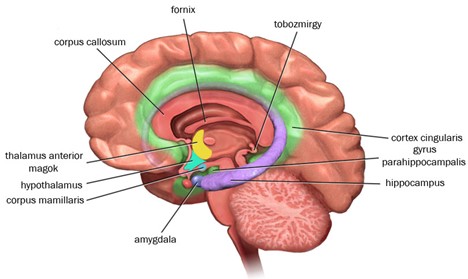
The Relationship between Emotions and Common Brain Structures Hippocampus
The hippocampus is involved in the creation of vivid, episodic memories. An episodic memory is a memory of a specific event or situation we have experienced – such as our first day of college! Episodic memories involve emotional content. In fact, the amygdala and hippocampus work together to encode episodic memories. The amygdala determines whether an event has a strong emotional component. If yes, then the amygdala tags the event as emotional and sends the information to the hippocampus for encoding.
Reproduced from “Limbicus rendszer” by Capucettorosso, 2019. Open Access, Creative Commons Attribution-Share Alike 4.0 International. Retrieved from File:Brodmann areas.jpg – Wikimedia Commons
One way to further identify the functions of brain structures is to evaluate the impact of damage to one structure on our emotional experiences. One study recruited wounded U.S. soldiers – one group who had damage outside the amygdala and another who had brain damage that included the amygdala (Koenigs et al., 2008). Those soldiers who experienced damage to the amygdala did not develop PTSD, while 40% of those soldiers who had no damage to the amygdala developed PTSD! Why? What does this have to do with the hippocampus? If our amygdala is damaged, then our amygdala cannot tag events as fearful or emotional so the amygdala does not send the information about the emotional event to the hippocampus for encoding.
In another study (Kalin & Shelton, 2000), young rhesus monkeys were exposed to three conditions and their behavioral responses were evaluated. Figure 8 shows drawing of these conditions. It is important to note that the monkeys were separated from their mothers, which elicits distress. After separation from the mother, the young monkeys were placed in a cage and experienced each condition for a three- to nine-minute period. In the first alone condition, the monkeys were left alone. In the alone condition, normal behavior included increased bodily movement and cooing, which the researchers believe is similar to crying to help the mother locate the infant monkey. In the second condition, the monkeys saw the profile of a human intruder, but the intruder never made direct eye contact with the monkeys. During this no-eye-contact profile condition, typically monkeys reduced cooing and increased freezing and/or hiding behavior. In the third condition, a human intruder stared at and made eye contact with the monkey. In this stare condition, the monkey either exhibited aggressive behavior (e.g., barking, shaking, and approaching/lunging) or submissive behaviors, labeled “lip smacking and fear-grimacing” (p. 52). Typically, monkeys showed more freezing behavior during the profile condition than the two other conditions. Out of 100 monkeys, three monkeys showed context-inappropriate responding – which occurs when the behavior changes do not match the context of the emotional event or when the behavior changes last long after the emotional event has ended. These 3 monkeys showed context-inappropriate responding by exhibiting freezing, not aggressive, behaviors in the stare condition. Further results showed these three monkeys had greater activation in the right cortex and elevated cortisol levels, changes associated with the emotion fear. How does this relate to the hippocampus? Well, researchers (Davidson et al., 2000; Kalin and Shelton, 2000) believe that the hippocampus helps us to switch our emotions based on changing situations – such as the situation changing from the profile to stare conditions for the monkeys. Monkeys with abnormally functioning hippocampi may have more trouble adjusting behaviors to new contexts. Further, increased cortisol may be associated with hippocampus abnormalities and the tendency to experience fear. It is important to note that Kalin and Shelton (2000) and others are theorizing that the hippocampus damage or size could be one explanation of context-inappropriate responding (which is a symptom of depression and anxiety disorders).
Figure 8
Experimental Conditions from Kalin and Shelton
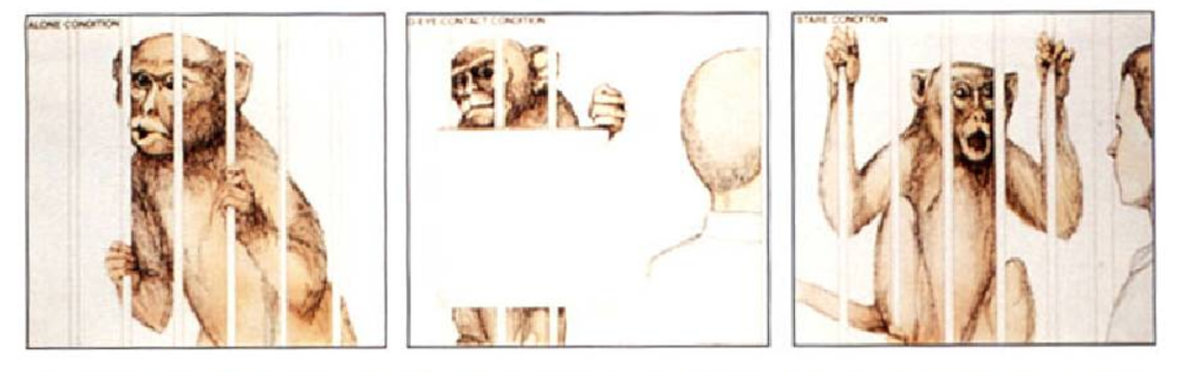
Note. In order from left to right, conditions are: 1) alone 2) profile – no eye contact and 3) stare – direct eye contact. Reproduced from “The regulation of defensive behaviors in Rhesus monkeys: Implications for understanding anxiety disorders,” by N.H. Kalin, and S.E. Shelton, 2000, In R. J. Davidson (Ed,), Anxiety, depression and emotion, p. 52. Copyright 2000 by Oxford University Press
