
Chapter 7: Physiological Measures of Emotion
Reward Circuit
The reward circuit is comprised of many structures, two of which are the basal ganglia and the ventral tegmental area (VTA; see Figure 10). The basal ganglia includes the ventral striatum (which encompasses the nucleus accumbens) and the dorsal striatum (which includes the caudate nucleus). The VTA makes and releases dopamine into the nucleus accumbens. Many of these structures are activated when seeking rewards like drugs, alcohol, and sexual intercourse, as well as associated with romantic love (for a review, see Haber & Knutson, 2010 and Ranaldi, 2014). These structures are activated when people are anticipating and thinking about a reward, but are not activated when people are actually consuming or enjoying the reward. Thus, these structures might motivate people to approach rewards. Activation of reward structures is positively correlated with the magnitude and likelihood of the anticipated reward (Knutson et al., 2001, 2005). The bigger the reward and the more likely one is to receive the reward – the greater the activation in these structures! More recent work has found that the reward system is activated for pride (Roth et al., 2014), gratitude, and when engaging in prosocial behaviors (Moll et al., 2006; Rilling et al., 2002). But, some work suggests this the reward system is activated for emotions like schaedenfraude (Takahashi et al., 2009), and negative emotions like shame (Roth et al., 2014). Clearly, these structures play an important role in positive emotions. Damage to these structures could make experiencing positive emotions harder, leading people to seek out riskier rewards such as drugs and video games to feel pleasure. Alternatively, damage might actually elicit dislike to events that initially caused positive emotions (for a review, Berridge & Kringelbach, 2008).
Figure 10
Reward System
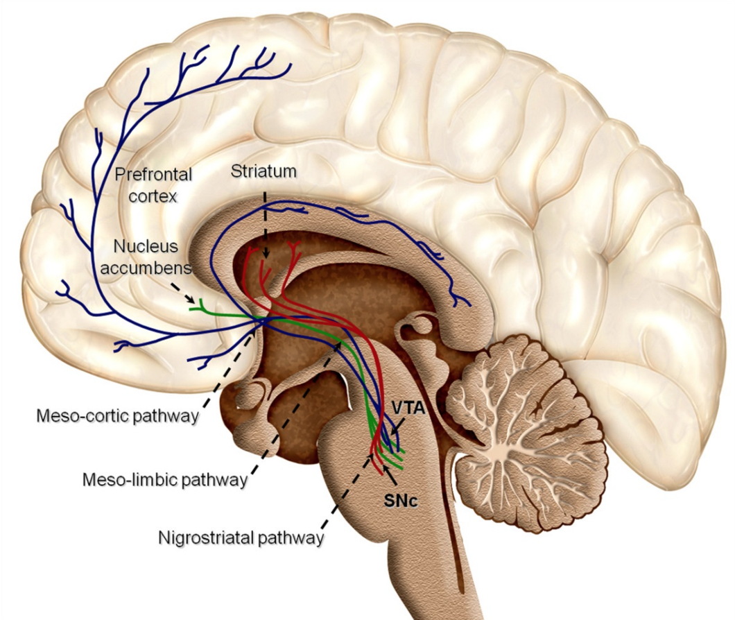
Reproduced from , “Dopaminergic reward system: A short integrative review” by O.C. Arias-Carrión, M.Stamelou, E. Murillo-Rodríguez, M. Menéndez-González, and E. Pöppel, 2010, International Archives of Medicine, 3(24), p. 2 (IntarchMed – International Archives of Medicine). Open Access. Creative Commons Attribution 3.0 Unported. Retrieved from: Dopaminergic reward system: a short integrative review
