
Chapter 7: Physiological Measures of Emotion
Physiological Measures of ANS Activity
This section will cover the ways that researchers measure ANS activity. Remember, it can be hard to determine whether both the SNS and PNS are working simultaneously or separately. For an excellent review of measurement tools used in emotions research, please read Mauss and Robinson (2009).
Measures of SNS or PNS Activity
Several physiological measures can measure ANS activity but are not considered pure measures of SNS or PNS activity. Some of these measures include blood pressure, respiration rate, and measures of pupil activity. Systolic blood pressure is blood pressure measured while the heartbeat is actively pushing blood through the arteries. Diastolic blood pressure measures blood flow while the heart is between beats. Respiration rate measures our breathing rate and includes measures of breathing rate and breathing depth. Pupil measures include gaze location and pupil size. During SNS activity, blood pressure and breathing increase and pupils dilate – but remember these measures are not pure measures of SNS activity.
Measures of SNS Activity Only
Physiological measures that provide pure measures of SNS activity include skin conductance level and cardiac pre-ejection period. Skin conductance level is not influenced by the PNS system. So, when skin conductance increases this indicates the SNS system is working. Skin conductance level measures the electrical conductance of the skin, which varies when we sweat. To measure skin conductance, two sensors are placed on the forefingers of each hand. One sensor sends an electrical signal through to the other finger and the system determines how long it takes the finger to reach the other finger. Skin resistance is the time it takes the electrical signal to travel from one sensor to the other. Skin conductance is calculated as the inverse of skin resistance (1/skin resistance).
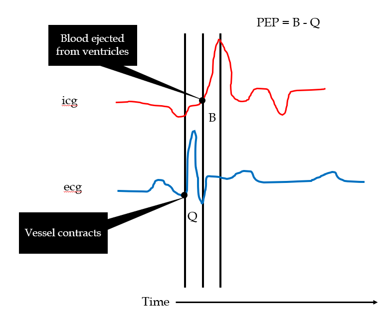
Cardiac pre-ejection period (PEP) is usually measured in combination with RSA to tweeze apart SNS and PNS activity. In Figure 2 on the EKG printout, the cardiac pre-ejection period is the time between when the heart vessel contracts (B) and the valves open and eject blood (Q). Eventually this blood is ejected through the aortic valve. PEP is calculated by subtracting Q from B (PEP = B – Q). Faster time periods (or fewer seconds) indicate greater activation of the SNS.
Figure 2
Cardiac PEP displayed on EKG printout
Measures of PNS Activity Only
Measures of pure PNS activity include heart rate variability and vagal tone, which are both related to each other. Before explaining these two measures, I first need to explain respiratory sinus arrhythmia (RSA). Arrhythmia means irregularity or to skip. Typically, heart arrhythmias are diagnosed irregularities in heartrate. But, RSA is a naturally occurring irregularity in the heart rate that occurs when we take a breath. Everyone has an RSA. When we inhale our heart rate increases, and when we exhale our heart rate decreases. To measure RSA, heart-rate variability is used.
Heart-rate variability (HRV) is calculated as our inhaling heartrate minus our exhaling heartrate. HRV indicates how strongly the PNS is working to slow down our heart rate while we breathe. A higher HRV number indicates activation of the PNS.
Respiratory Sinus Arrhythmia
Vagal tone measures the degree of PNS activity on the heart while a person is at rest. The vagus nerve (see Figure 3) operates during PNS activity and specifically communicates between internal organ functioning and the brain. Vagal tone measures vagus nerve activity during rest and digestion, but currently there is not a direct method to measure vagal tone activity. As a work around, researchers measure heart rate and heart-rate variability because the vagus nerve impacts the function of the internal organs like the heart and stomach.
Figure 3
Vagus nerve (in yellow)
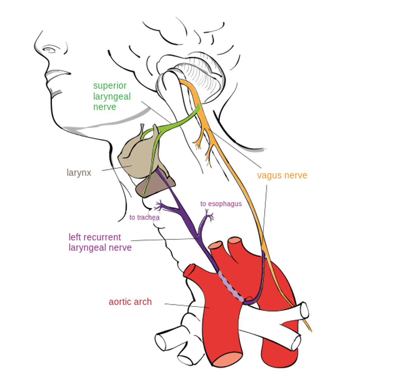
Reproduced from “Drawing of the left recurrent laryngeal nerve” by Jkwchui, 2014. Open Access, Creative Commons Attribution-Share Alike 3.0 Unported. Retrieved from: File:Recurrent laryngeal nerve.svg – Wikimedia Commons
Watch Dachnar Keltner Explain the Vagus Nerve
Increased vagal tone (which means the vagus nerve is currently operating) is associated with a reduced heart rate and a more variable heart rate (i.e., higher HRV). HRV, vagal tone, and PNS activity are positively correlated. High HRV and high vagal tone are associated with better emotion regulation, and the tendencies to experience positive emotions and relationship-oriented emotions like compassion. New work is suggesting that reduced or less complex heart rate variability during REM sleep could be a predictor of major depression (Kwon et al., 2019).
