
Chapter 8: Fear, Anxiety, and Stress
Fear vs. Anxiety: Physiological and Brain Differences
This section will highlight some of the physiological differences between fear and anxiety that can help us to further differentiate between these two constructs.
Startle Reflexes. Anxiety and fear both result in startle reflexes, but these reflexes are different. In a fear response, people experience a startle response to an unexpected threat like an unexpected sound or light. This is called the fear-potentiated startle test (Davis et al., 1997). This fear startle response is quick to dissipate because the unexpected event quickly stops. Conversely, during anxiety, animals experience a prolonged startle response that does not immediately dissipate. This prolonged response has been demonstrated in rats who stare at a bright light for 5 to 20 minutes (Walker & Davis, 1997). This is called the light-enhanced startle effect (Davis et al., 1997). The bright light does not immediately cause the startle reflex, but the prolonged stimulus results in a slow-onset and long startle reflex. Unlike the fear reflex, this startle response remains even after the light is turned off. During a specific threat, people diagnosed with fear disorders show a greater increase in heart rate and greater startle reflex than participants diagnosed with an anxiety disorder. During baseline (when an eliciting event is not present), people diagnosed with anxiety disorders show an increase in heartrate and increased startle reflex compared to people with fear disorders.
Brain Activation. Fear and anxiety demonstrate activation in different structures of the brain. Fear activates the central nucleus of the amygdala, whereas anxiety activates the bed nucleus of the stria terminalis (BNST), a structure located near the amygdala (see Figure 18 below; Davis et al., 1997). The central nucleus of the amygdala is activated for responses to specific fear eliciting events and is activated by the fear-potentiated startle test. Conversely, the bed nucleus of the stria terminalis (BNST) is part of the extended amygdala. Here is a link for more information.
The BNST is activated without a specific stimulus and is activated by more long-lasting responses to negative emotional stimuli.
Figure 18
Location of Amygdala and the BNST
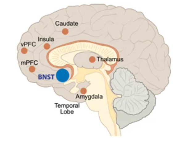
Reproduced from “Over-shadowed by the amygdala: The bed nucleus of the stria terminalis emerges as a key to psychiatric disorders,” by M.A. Lebow and A. Chen, 2016, Molecular Psychiatry, 21, p. 455. (Overshadowed by the amygdala: the bed nucleus of the stria terminalis emerges as key to psychiatric disorders). Retrieved from: Overshadowed by the amygdala: the bed nucleus of the stria terminalis emerges as key to psychiatric disorders. Copyright 2016 by Macmillan Publishers Limited, Open Access.
Figure 19 below shows the fear and anxiety brain networks, both which lead to similar structures listed in the center (e.g., lateral hypothalamus, reticular formation). Further, note that the BNST and amygdala are directly connected and may send information to each other. CRH stands for Corticotropin-releasing hormone. CRH is a stress peptide which regulates the release of cortisol during a stressful, but not a fear-provoking, event. As exemplified in Figure 19, during the prolonged startle reflex, CRH acts on the receptors in the BNST, but not the central nucleus of the amygdala. The bed nucleus is activated for a longer lasting startle reflex that typically releases cortisol, instead of a quick startle associated with fear. When the BNST is lesioned in rats and mice, the fear startle still occurs, but the longer-lasting anxiety startle reflex cannot occur (Davis et al., 1997). Conversely, when the central nucleus of the amygdala is lesioned, the prolonged anxiety startle can occur, but the lesions prevents the short fear startle.
Figure 19
Depiction of brain structures and processes involved in fear and anxiety
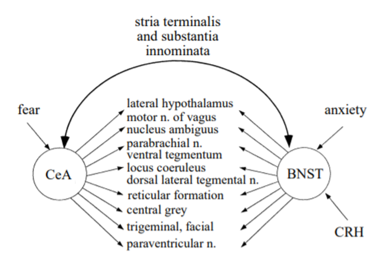
Note. CeA = central nucleus of the amygdala, BNST = bed nucleus of the stria terminalis, CRH = Corticotropin-releasing hormone. Reproduced from “Amygdala and bed nucleus of the stria terminalis: differential roles in fear and anxiety measured with the acoustic startle reflex,” by M. Davis, D.L. Walker, and Y. Lee, 1997, Philosophical Transactions of the Royal Society of London. Series B: Biological Sciences, 352(1362), p. 1685 (Amygdala and bed nucleus of the stria terminalis: differential roles in fear and anxiety measured with the acoustic startle reflex). Copyright 1997 by The Royal Society.
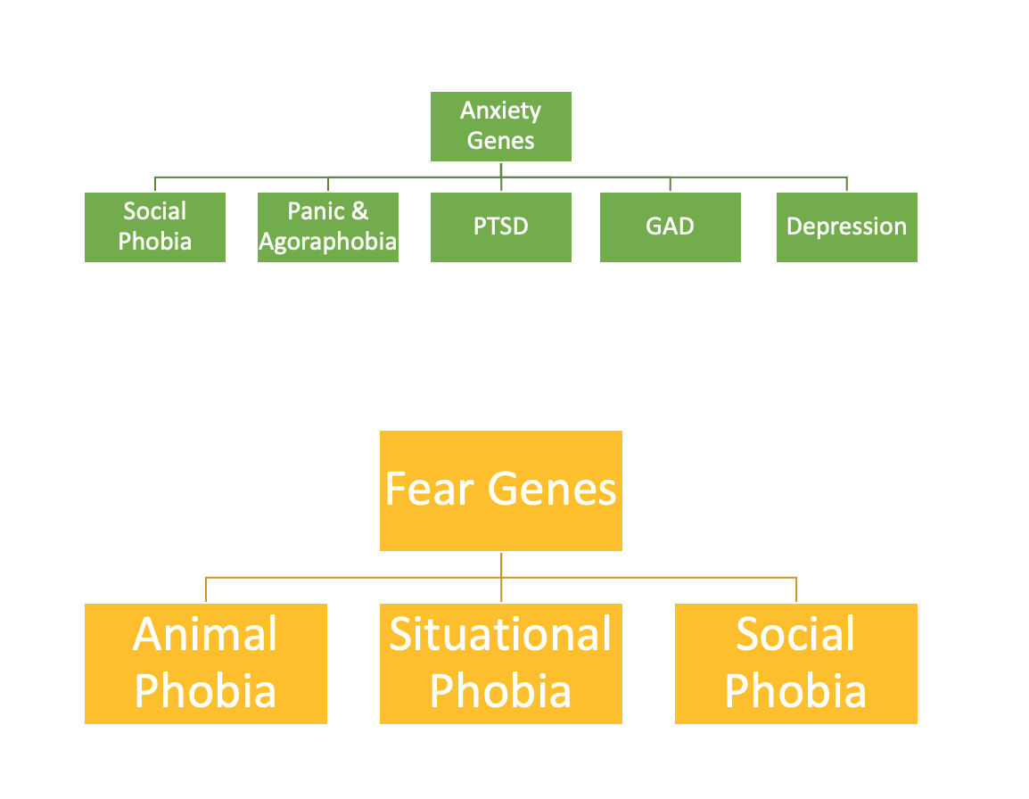
Figure 20
Genetic Structure of Fear and Anxiety Disorders
Yale Experts in Emotion video on Anxiety and Emotion with Dr. Douglas Mennin
