How many dimensions underlie physiological measures?
Russell (1980) and Watson and Tellegen (1985) showed subjective feelings can be reduced to a small number of dimensions. But what about other emotion components like physiological changes and behavior changes?
Lang et al. (1993) investigated whether subjective feelings, facial behavior changes, and arousal could together be reduced to a small number of dimensions. Participants viewed a variety of photos from the Internal Affective Picture System (IAPS; more on IAPS later!). Participants viewed 18 different photos: 8 photos were pleasant in valence (nature, food, attractive people), 8 photos were unpleasant (pollution, accidents, mutilated bodies) and 2 photos were neutral (household objects). While viewing the photos, participants self-reported valence, arousal, and interest in the photos. In addition, the amount of time participants looked at each picture was recorded. While viewing the photos, researchers measured facial electromyographic (EMG) activity and physiological measures (i.e., heart rate, skin conductance, startle reflex). Facial EMG measures changes in the facial muscles, such as the corrugator and zygomatic muscles (see Figure 8). Corrugator facial activity is correlated with valence, such that increases in activity is associated with negative emotions and decreases are associated with positive emotions. The zygomatic muscle is the muscle around the mouth used to smile. Increases in zygomatic activity suggests a positive emotional experience. Researchers then correlated participants’ self-reported valence and arousal with changes in facial muscles and physiology. It is important to note that higher numbers on the self-report scale indicate higher levels of pleasantness or arousal.
Figure 8
Location of Corrugator and Zygomatic Facial Muscles
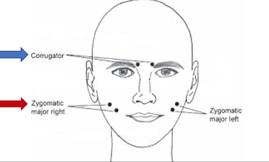
Table 2
Lang et al. (1993) Results
|
Corrugator Activity
Negatively correlated with self-reported pleasant valence
No relationship with self-reported arousal
|
|
Zygomatic Activity
U-shapred relationship with self-reported valence
Positively correlated with self-reported arousal
|
|
Heart Rate
Positively correlated with self-reported pleasant valence
Positively correlated with self-reported arousal
|
|
Skin Conductance
Positively correlated with arousal
No relationship with valence
|
|
Startle Reflex
Negatively correlated with self-reported pleasant valence
No relationship with arousal
|
After finding the results (see Table 2), researchers conducted factor analysis on all data (self-report, facial EMG, physiology). Remember, factor analysis is a statistical analysis that reduces all data into a smaller number of groups. So, this study is reducing subjective feelings, behavior change, and physiology into smaller groups. Factor analysis returned two clear factors – valence and arousal (see Figure 9 below)! The first group was valence. Pleasant valence was associated with an increase in self-reported pleasantness, decrease in corrugator activity, increase in zygomatic activity, increase in heart rate, and decrease in startle reflex. (Later we will discuss how the startle reflex is a pure measure of valence, such that the startle reflex is positively correlated with unpleasantness). On the second factor, high levels of arousal were associated with an increase in self-reported arousal and interest, greater viewing time, and higher skin conductance.
Two Underlying Dimensions from Lang et al. (1993)
[Lang, P.J., Greenwald, M.K., Bradley, M.M., & Hamm, A.O. (1993). Looking at pictures: Affective,facial, visceral, and behavioral reactions. Psychophysiology, 30, 261-273]
| Pleasant Valence |
High Arousal |
- Self-reported pleasant valence
- Corrugator (-)
- Zygomatic
- Heart Rate
- Startle Reflex (-)
|
- Self-reported arousal
- Self-reported interest
- Viewing Time
- Skin Conductance
|
Across several studies conducted by Russell, Barrett, and others (Colibazzi et al., 2010; Posner et al., 2009; Wilson-Mendenhall et al., 2013) researchers replicated Lang et al.’s (1993) study by measuring changes in brain activity. In this study, participants rated the valence and arousal in three tasks: 1) emotion words, 2) sentences with emotional content, and 3) emotional scenarios. While reading and rating these tasks, participants brain activity was measured. Again, these researchers found two underlying dimensions – valence and arousal! (see Figure 10). In general, results showed that activation of the insular cortex, reward circuit (i.e., prefrontal cortex, basal ganglia, ventral striatum, nucleus accumbens, and ventral tegmental area (VTA)), and orbitofrontal cortex was associated with self-reported pleasantness. Activation of the thalamus, amygdala, parahippocampal gyrus, and anterior cingulate was associated with self-reported arousal. It is important to note that the orbitofrontal cortex is also activated for anger (a negative, approach emotion), the insular cortex with disgust and amusement, and that the anterior cingulate cortex is also linked to sadness (a low arousal, negative emotion). Thus, there may not be clear one-to-one mappings of these structures to valence and arousal.
Two Underlying Dimensions from Colibazzi et al. (2010), Posner et al. (2009) and Wilson-Mendenhall et al. (2013)
[Wilson-Mendenhall, Barret, & Barsalou, 2013; Posner, Russell…Colibazzi, et al., 2009; Colibazzi, Posner,…. Russell, 2010]
| Pleasant Valence |
High Arousal |
- Self-reported valence
- Insular Cortex
- Reward Circuit
- Orbitofrontal cortex
|
- Self-reported arousal
- Thalamus
- Amygdala
- Parahippocampalgyrus
- Anterior cingulate
|

