
Chapter 7: Physiological Measures of Emotion
One-Network Hypothesis
The one-network hypothesis comes from the social constructivist perspective. This hypothesis points out that an emotional experience is comprised of behavior, thoughts, feelings, and physiology, all which activate different parts and structures of the brain. Theorists who support this perspective point out that one emotion can activate several parts of the brain and that brain structures cannot be linked uniquely to one structure. This, theory seeks to find networks or connections of brain structures that can be mapped onto emotion components, not specific emotions. Some hypothesized networks include core affect, conceptualization, executive attention, and emotion words (Lindquist et al., 2012). Core affect , as discussed with Russell’s (1980) circumplex model, is a general feeling of pleasantness or unpleasantness accompanied by some feeling of arousal. Conceptualization essentially represents our cognitive appraisal of the emotional event. Executive attention is activated when we are attending to the emotional event and focusing on certain aspects of the event, while ignoring other aspects of the event. The emotion words network is activated when we consciously assess our core affect, behaviors, facial expressions, and other components to determine an emotion word label. These are just some of the hypothesized networks.Figures 12 and 13 display some emotional networks supported by past research (for a review, see Lindquist et al., 2012). Core affect includes the amygdala, insula, orbitrofrontal cortex, anterior cingulate cortex, strai terminalis, and PAG. Conceptualization includes areas such ventromedial prefrontal cortex (VMPFC) and hippocampus.
Figure 12
Display of Unique Emotional Networks

Reproduced from “The brain basis of emotion: a meta-analytic review,” by K.A. Lindquist, T.D. Wager, and H. Kober, E. Bliss-Moreau, and L.F. Barrett, 2012, The Behavioral and brain sciences, 35(3), p. 55 (The brain basis of emotion: A meta-analytic review). Copyright 2011 by the Cambridge University Press.
Figure 13
Display of Emotional Networks from Different Views of the Brain
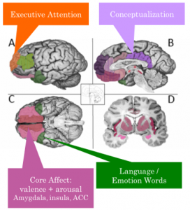
Reproduced from “The brain basis of emotion: a meta-analytic review,” by K.A. Lindquist, T.D. Wager, and H. Kober, E. Bliss-Moreau, and L.F. Barrett, 2012, The Behavioral and brain sciences, 35(3), p. 54 (The brain basis of emotion: A meta-analytic review). Copyright 2011 by the Cambridge University Press.
