
Chapter 12: Pride – A Positive Self-Conscious Emotion
Physiological Changes
Table 5
Physiological Changes Caused by Authentic Pride
| How Pride was Elicited in Study | Physiological Changes |
|---|---|
| Film Clips | Increased HR, Increased SCR, 0 HRV |
| Recall Pride Experience | Increased SCR |
| Experimenter Praise | Increased HR, 0 Cardiac PEP |
Adapted from “Autonomic nervous system activity in emotion: A review,” by S.D. Kreibig, 2010, Biological Psychology, 84(3), p. 407 (https://doi.org/10.1016/j.biopsycho.2010.03.010) Copyright 2010 by Elsevier B.V.
Two studies investigated the brain structures activated for pride experiences. In the first study (Roth et al., 2014), participants completed three conditions in which guilt/shame, pride, and neutral emotions were elicited. To elicit each emotion, participants were asked to recall a time between 3 weeks and 6 months ago that they felt ashamed/guilty, a time they felt pride, and for the neutral condition were asked to simply wait. The procedures are displayed in Figure 15 below. Participants watched a screen and were given a cue that indicated which of the three conditions to think about. During the task step, participants were either thinking about the emotion episode or waiting in the neutral condition. Then, participants viewed a neutral IAPS image to help their physiology return to baseline. In the fourth step, baseline measures were taken prior to starting one of the three conditions. During these four steps, an fMRI machine is measuring brain activation. Table 6 displays the findings. In this study, the two emotion conditions versus the neutral condition showed greater activation in several structures. The authors note that a possible emotion network might be the amygdala, insula, and ventral striatum. This emotion network may simply be activated when we experience any emotion. The MPFC and the PCC are activated when we think about and evaluate the self and thus would be activated for negative and positive self-conscious emotions. Pride showed more activation than shame in five different structures, but we do not yet know what these findings suggest.
Figure 15
Example of Trials from Roth et al. (2014)
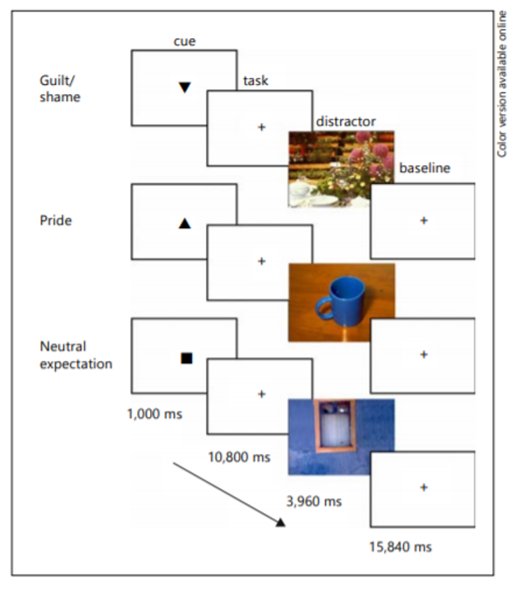
Reproduced from “Brain activation associated with pride and shame,” by L. Roth, T. Kaffenberger, U. Herwig, and A.B. Brühl, 2014, Neuropsychobiology, 69(2), p. 96 (https://doi.org/10.1159/000358090) Open Access, Zurich Open Repository and Archive, University of Zurich.
Table 6
Summary of Roth et al. (2014) Findings
| Comparison | Findings |
|---|---|
| Pride and Shame/Guilt (vs. Neutral) |
|
|
|
| Shame/Guilt (vs. Pride) |
|
Adapted from “Brain activation associated with pride and shame,” by L. Roth, T. Kaffenberger, U. Herwig, and A.B. Brühl, 2014, Neuropsychobiology, 69(2), p. 98-101 (https://doi.org/10.1159/000358090) Open Access, Zurich Open Repository and Archive, University of Zurich.
Table 7
Summary of Takashashi et al. (2008) Findings
| Comparison | Findings |
|---|---|
| Pride (vs. Neutral) |
|
| Joy (vs. Neutral) |
|
| Pride (vs. Joy) |
|
Adapted from “Brain activations during judgments of positive self-conscious emotion and positive basic emotion: pride and joy,” by H. Takahashi, M. Matsuura, M. Koeda, N. Yahata, T. Suhara, T., M. Kato, M., and Y. Okubo (2008). Cerebral Cortex, 18(4), p. 900 (https://doi.org/10.1093/cercor/bhm120) Copyright 2007 by The Author.
