
Chapter 8: Fear, Anxiety, and Stress
Amygdala and Fear: Klüver-Bucy Syndrome and Urbach-Weithe Disease
In this section, we will discuss the amygdala’s role in the emotion fear. The human brain includes two amygdalae, both of which are located in the temporal lobe. The amygdala is often mentioned as one of the brain structures that is activated during a fearful experience. In this section, we will expand by investigating how the amygdala is related to other emotions.
Figure 10
Amygdalae in Red Viewed from Side (left) and Back (right) of Brain
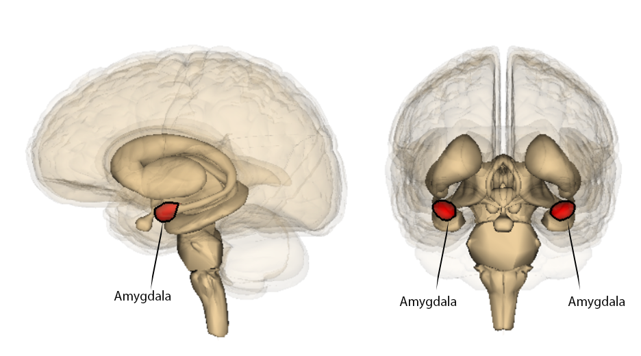
Reproduced from “Amygdala” by Life Science Database (LSDB), 2009. Open Access, Creative Commons Attribution-Share Alike 2.1 Japan, CC-BY-SA-2.1-jp. Retrieved from: File:Amygdala.png
Long Description
The image displays two anatomical views of the human brain, each highlighting the amygdala in red:
- Left Side: A sagittal view (side profile of the brain) shows the location of the amygdala deep within the temporal lobe. The structure is clearly marked and labeled in red.
- Right Side: A coronal view (front-facing cross-section) shows both the left and right amygdalae, symmetrically positioned and colored red. Each is labeled to indicate its location within the medial temporal lobes.
This image is typically used in neuroscience or psychology to illustrate the position of the amygdala, a brain region involved in emotion processing, especially fear and threat detection.
One way to evaluate the role of the amygdala in emotions is to conduct lesion studies. A lesion study investigates how damage in the amygdala effects people’s emotional experiences compared to a control group without damage. One way to conduct a lesion study would be to damage the amygdala in rats and assess how that impacts emotion. A second way would be to recruit humans who have damage to the amygdala. Klüver-Bucy Syndrome (Klüver & Bucy, 1939) describes a variety of symptoms associated with amygdala damage. Klüver-Bucy can be caused by stroke, encephalitis, tumors, and even a lobotomy. Typically, Klüver-Bucy is expressed when one or both amygdalae, and often parts of the temporal lobe, have been damaged. In 1939, Klüver and Bucy first described the symptoms after conducting bilateral temporal lobectomies in rhesus monkeys. After the lobectomy, monkeys showed odd oral behaviors such as sticking objects in their mouths that they typically do not. The monkeys showed hypersexual behaviors, such as efforts to mate with inanimate objects. A final symptom was that monkeys who were originally fearful and aggressive became placid when presented with fearful objects such as snakes. The removal of the amygdalae seemed to change emotional experiences such as reducing disgust elicited by placing harmful objects in the mouth, and lack of fear or aggression when presented with danger. These symptoms have been replicated with cats and even in humans. Humans who present with Klüver-Bucy show dampened emotions or even exaggerated rage and aggression. One thing to note is that Klüver-Bucy is often caused by damage to the amygdala and surrounding areas, thus any changes in emotions cannot be solely due to damage in the amygdala. Urbach-Weithe Disease is a rare genetic disorder caused by calcium build-up in the amygdala, which causes the brain tissue to harden. In Urbach-Weithe Disease, the damage typically only affects the amygdala – thus allowing researchers to link the symptoms to a specific structure in the brain. Urbach-Weithe symptoms are similar to Klüver-Bucy and include reduced fear, heightened aggression, and changes in the emotion of disgust.
