Chapter 8: Fear, Anxiety, and Stress
Evolutionary Evidence – Backward Masking and Nonconscious Processing of Threatening Stimuli
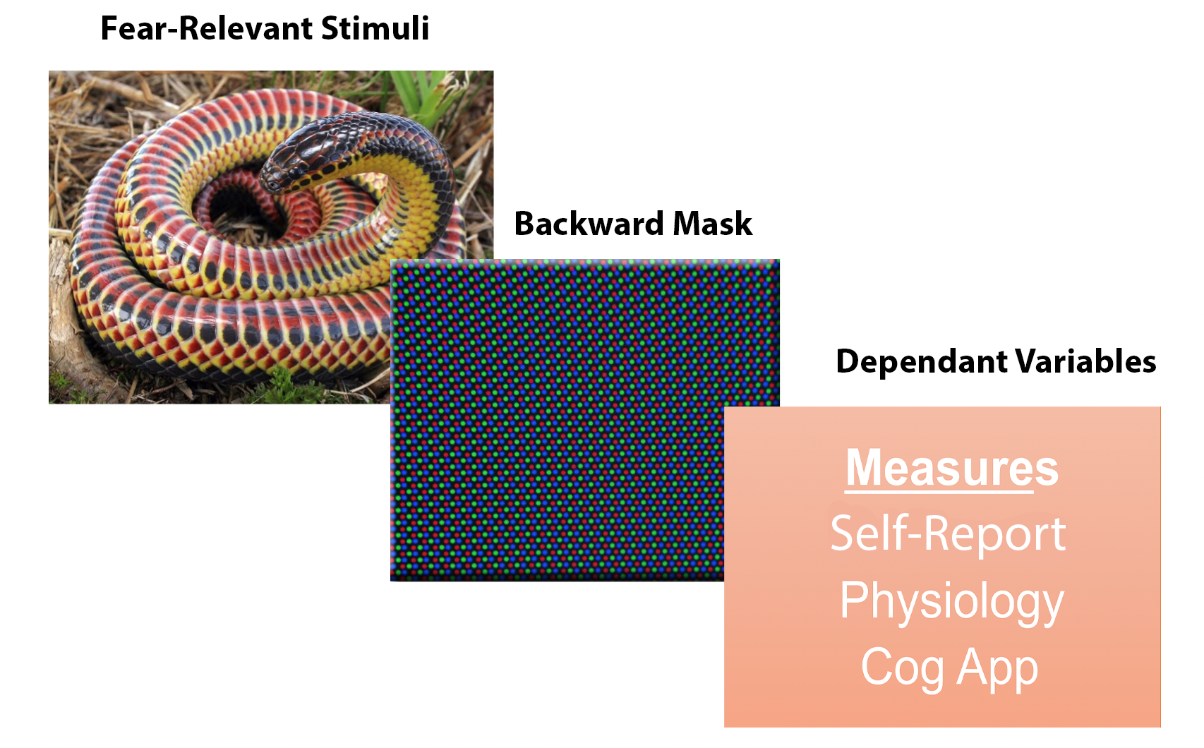
Figure 7
Example of Backward Masking Methodology

Snake image reproduced from “Farancia erytrogramma (rainbow snake)” by Charles Baker, 2018. Open Access, Creative Commons Attribution-Share Alike 4.0 International Retrieved from: File:Farancia erytrogramma (rainbow snake).jpg
Pixels image reproduced from “CRT pixel array” by w:User:Planemad, 2007. Open Access, Creative Commons Attribution-Share Alike 2.5 Generic. Retrieved from:
File:CRT pixel array.jpg
One study (Öhman & Soares, 1994) investigated fear with a backward masking procedure. The between-subjects quasi-IV was whether participants self-reported extreme fears of only snakes, only spiders, or neither (control). Within-subjects variables were target of photo (snakes, spiders, mushrooms, flowers), phase of image presentation (masked vs. unmasked) and number of trials (8 total trials). During each of the masked and unmasked phases, participants completed 8 viewing trials of each photo. So, participants viewed 8 masked photos of snakes, 8 masked photos of spiders, and so on. The dependent variables were skin conductance (a pure measures of SNS activity), and self-reported valence, arousal, and dominance on the Self-Assessment Mannikin (SAM; Bradley & Lang, 1994). While viewing these photos, skin conductance was measured. In the masked phases, the target photos were shown for 30 milliseconds, then followed by the mask for a period of 100 milliseconds. In the unmasked phases, the images were shown for 130 milliseconds. After completion of each of the masked and unmasked phases, participants were shown the four images again (masked or unmasked based on phase just completed) from the prior phase and reported the following: 1) image they saw in the photo, and 2) self-reported arousal, valence, and control-dominance to each picture. (Control-dominance could be considered a cognitive appraisal, similar to perceptions of control). Overall, most participants could not consciously identify the masked image (demonstrating the mask worked), while most could identify the unmasked image.
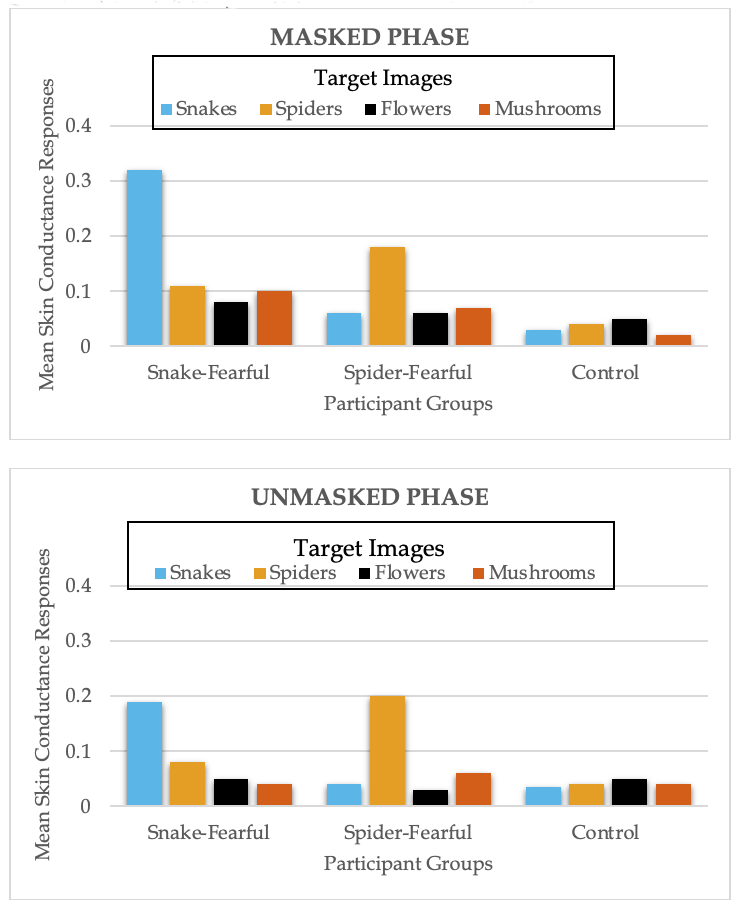
Figure 8 displays the results from the masked and unmasked phases. The results were the same for both masked and unmasked phases. The first result is that collapsed across participants groups, participants showed greater skin conductance to images of snakes and spiders compared to control images of flowers and mushrooms. This might suggest an evolutionary preparedness to experience fear toward threatening stimuli, even when the threatening stimuli are processed at the nonconscious level. The interaction between image condition and participant group was significant. When viewing the snake images, snake-fearful participants, compared to the other two groups, exhibited the greatest skin conductance. Similarly, when viewing spider images, spider-fearful participants showed the greatest skin conductance, compared to snake-fear and control participants. The three participant groups did not show differences in skin conductance when viewing flower or mushroom images. These findings suggest that physiological changes associated with fear occur at a preattentive level – prior to the participants being consciously aware of the threatening stimuli.
Figure 8
Skin Conductance Responses in Masked and Unmasked Phases

Long Description
The image features two bar graphs comparing mean skin conductance responses to four types of target images—snakes, spiders, flowers, and mushrooms—across three participant groups: Snake-Fearful, Spider-Fearful, and Control. The graphs are labeled:
- Masked Phase (top graph)
- Unmasked Phase (bottom graph)
Each graph has:
- Y-axis: Mean skin conductance response (ranging from 0 to 0.4).
- X-axis: Participant groups (Snake-Fearful, Spider-Fearful, Control).
- Bars: Color-coded by target image — Snakes (blue), Spiders (orange), Flowers (black), Mushrooms (yellow).
Masked Phase Highlights:
- Snake-Fearful: Strong response to snakes (~0.33), minimal to others.
- Spider-Fearful: Strong response to spiders (~0.25), moderate to mushrooms (~0.2), low to others.
- Control: Low responses across all categories (~0.05).
Unmasked Phase Highlights:
- Snake-Fearful: Moderate response to snakes (~0.2), low to others.
- Spider-Fearful: High response to spiders (>~0.3), moderate to mushrooms (~0.15), low to others.
- Control: Uniformly low responses (~0.05).
These graphs likely illustrate how fear-related stimuli elicit stronger physiological responses, especially when unmasked.
Adapted from “”Unconscious anxiety”: Phobic responses to masked stimuli,” by A. Öhman, and J.J. Soares, 1994, Journal of Abnormal Psychology, 103(2), p. 236. (“Unconscious anxiety”: Phobic responses to masked stimuli.). Copyright 1994 by American Psychological Association.
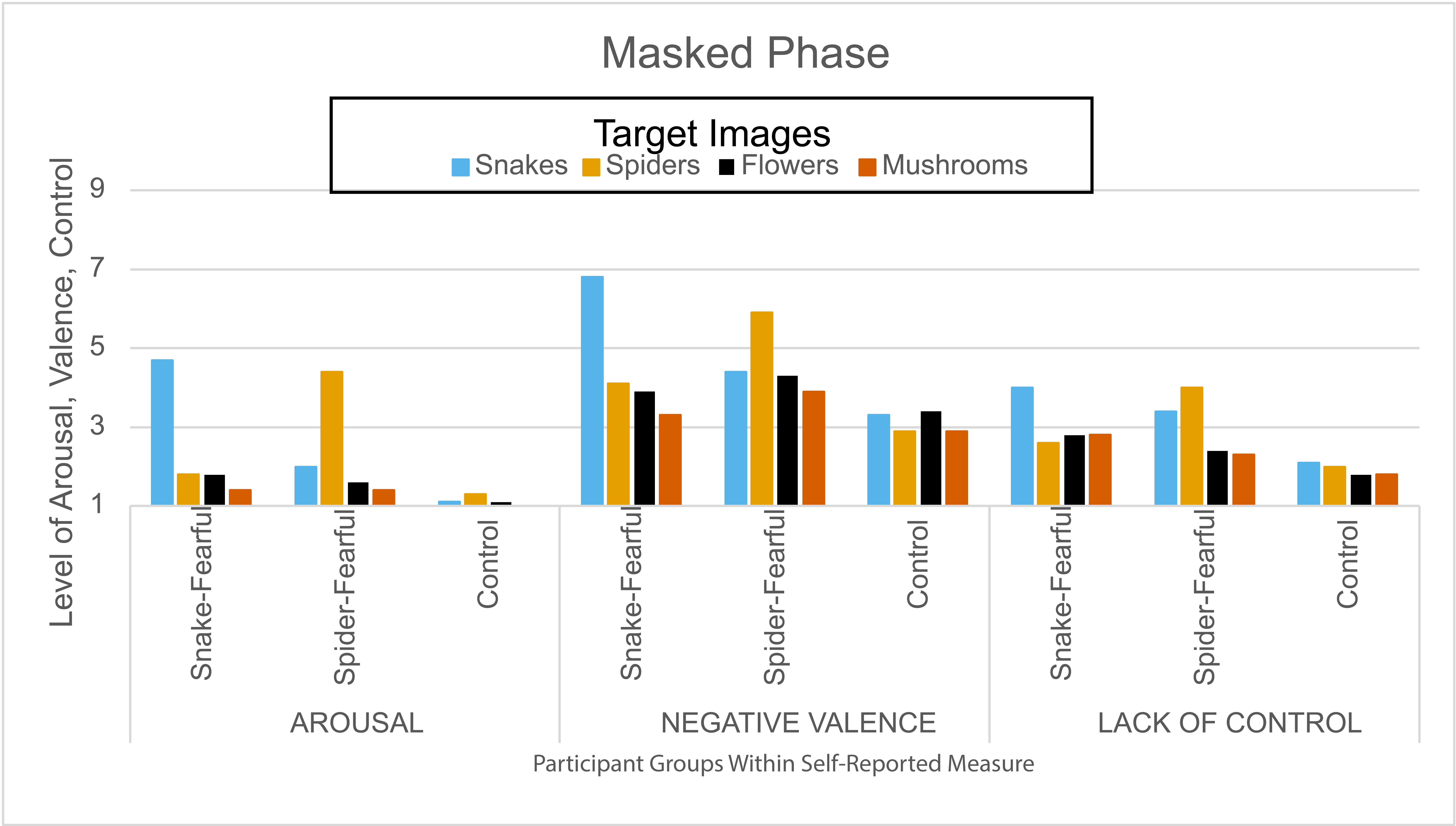
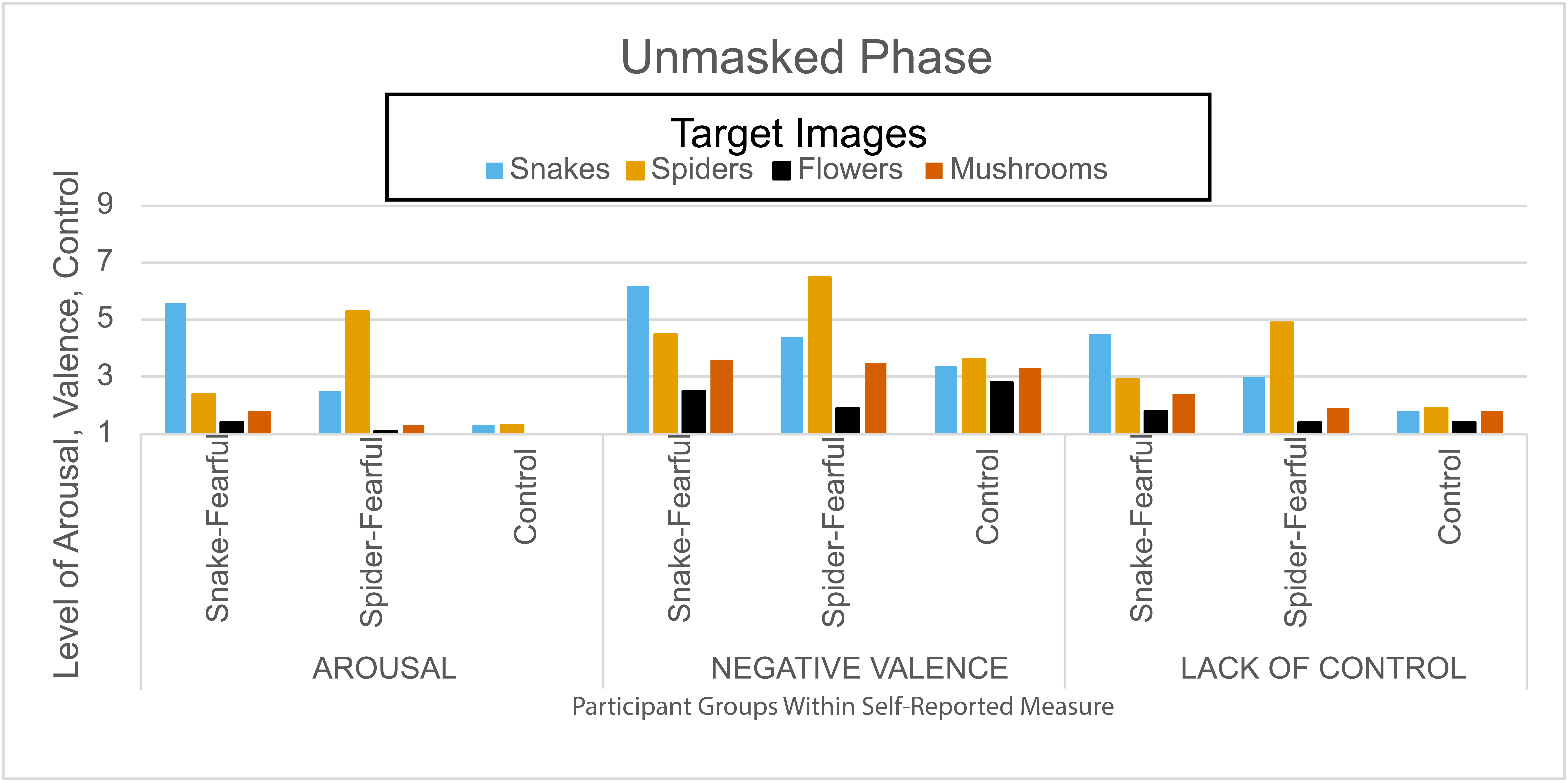
Figure 9
SAM Responses in Masked (Top) and Unmasked (Bottom) Phases

Long Description
The image is a single bar graph titled “Masked Phase”, displaying self-reported measures of emotional response across three psychological dimensions:
- Arousal
- Negative Valence
- Lack of Control
Axes:
- X-axis: Divided into the three dimensions above. Each dimension includes three participant groups:
- Snake-Fearful
- Spider-Fearful
- Control
- Y-axis: Measures response levels from 0 to 9.
Bar Colors (Target Images):
- Blue: Snakes
- Orange: Spiders
- Black: Flowers
- Brown: Mushrooms
Each participant group within each dimension has four bars representing their responses to the four image types. The graph visually compares how different groups perceive emotional arousal, negativity, and control when exposed to various stimuli under masked conditions

Long Description
The image is a bar graph titled “Unmasked Phase”, presenting self-reported emotional responses across three psychological dimensions:
- Arousal
- Negative Valence
- Lack of Control
Axes:
- X-axis: Divided into the three dimensions above. Each dimension includes three participant groups:
- Snake-Fearful
- Spider-Fearful
- Control
- Y-axis: Measures response levels from 1 to 9.
Bar Colors (Target Images):
- Blue: Snakes
- Orange: Spiders
- Black: Flowers
- Yellow: Mushrooms
Each participant group within each dimension has four bars representing their responses to the four image types. The graph visually compares how different groups perceive emotional arousal, negativity, and control when exposed to various stimuli during the unmasked condition of the study.
Adapted from “”Unconscious anxiety”: Phobic responses to masked stimuli,” by A. Öhman, and J.J. Soares, 1994, Journal of Abnormal Psychology, 103(2), p. 236. (“Unconscious anxiety”: Phobic responses to masked stimuli.). Copyright 1994 by American Psychological Association.
These findings show that when people are exposed to stimuli they already fear at a nonconscious level, they experience changes in physiology, subjective feelings, and cognitive appraisals. These findings further suggest that everyone does not universally experience fear to the same objects, which might contradict evolutionary theory. Current thought is that everyone has an evolutionary adaptation that prepares them to acquire fears to certain eliciting events, but the fears we acquire are going to depend on a combination of our genetics and past experiences with the threat. This view would suggest that people are not born with fears of snakes and spiders, but that we are born the with capacity to learn to fear certain events.
Further work supports the idea that the brain preattentively processes threatening stimuli. Patients with V1 or occipital lobe lesions experience a condition called blindsight. Blindsight occurs when a person does not consciously see a stimulus in their blind spot but can still identify the location and provide some information about the image. Patients with blindsight and complete blindness due to occipital lobe damage have shown greater right amygdala activation to fearful facial expressions compared to neutral expressions (Morris et al., 2001; Pegna et al., 2005). These findings provide further support to the idea that the amygdala can unconsciously process threatening stimuli, even though we cannot consciously see and process the stimuli.

