
Chapter 7: Physiological Measures of Emotion
Prefrontal Cortex
The functions of the prefrontal cortex (PFC) include planning, working memory, and inhibition of impulses. Related to emotions, the PFC is activated when people use emotional information to make decisions. For instance, people might think about how they will feel if they lie to their romantic partner (guilty!) and this prediction of the emotion guilt might help people to decide to not lie to their partner. The ventromedial prefrontal cortex (VMPFC) is a structure within the PFC. The VMPFC operates when people are anticipating how they will feel if they receive a reward (not how they will feel when they actually win the reward). The VMPFC is activated for taste, smells, touch, and social approval. When the PFC or VMPFC are damaged, people select the wrong outcomes in risky games because they cannot anticipate their emotions. Even though people understand and can verbally state the consequences of their actions, they have impaired ability to make decisions based on how they will feel after the action (e.g., sad, guilty, joyful, disappointed). Further symptoms include flat emotions, less empathy, and less reasoning about choices.
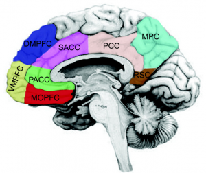
Reproduced from “Cortical midlines structures” by Georg Northoff, 2013. Open Access, Creative Commons Attribution 3.0 Unported. Retrieved from File:Brodmann areas.jpg – Wikimedia Commons
Long Description
This image is a labeled diagram of the human brain, highlighting various regions with distinct colors. Each label corresponds to a specific brain area, often referenced in neuroscience and cognitive research:
- DMPFC (blue): Dorsomedial Prefrontal Cortex – involved in social cognition and self-referential thinking.
- SACC (purple): Subgenual Anterior Cingulate Cortex – associated with mood regulation and emotional processing.
- PCC (pink): Posterior Cingulate Cortex – plays a role in memory and visual processing.
- MPC (green): Medial Parietal Cortex – involved in spatial orientation and navigation.
- RSC (brown): Retrosplenial Cortex – important for memory and navigation.
- PACC (light green): Perigenual Anterior Cingulate Cortex – linked to emotional and cognitive integration.
- VMPFC (yellow-green): Ventromedial Prefrontal Cortex – associated with decision-making and emotional regulation.
- MOPFC (red): Medial Orbitofrontal Prefrontal Cortex – involved in reward processing and value-based decision-making.
The diagram is likely used to illustrate functional brain networks or regions of interest in neuroimaging studies.
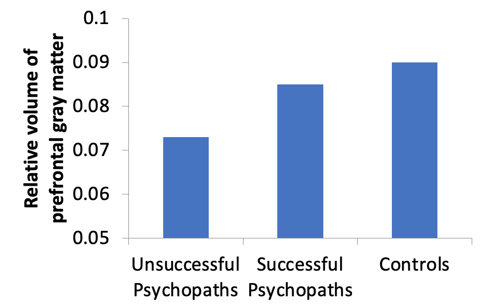
Studies have linked PFC activation and damage to the occurrence of crime. Researchers (Raine et al., 1998) divided criminals into two groups – 1) people who committed impulsive, emotional murders and 2) people who committed well-thought out and premeditated murders. Impulsive murders showed greater amygdala activation and reduced PFC activation These findings might suggest that impulsive murders experienced highly intense negative emotions (due to the amygdala), but reduced inability to inhibit the amygdala’s activity and control their emotions (because of the PFC). Another study (Yang et al., 2005) compared successful and unsuccessful male psychopaths (all determined to be psychopaths from a diagnostic checklist). Successful psychopaths were males who self-reported high levels of crime, but who had not been caught for their crimes. Conversely, unsuccessful psychopaths were convicted for their criminal acts. A third group of participants represented the control group and did not meet the requirements for psychopathy. Results showed that higher psychopathy scores were linked to lower volume of prefrontal gray matter. In fact, as shown in Figure 9, unsuccessful psychopaths (compared to male controls) showed a 22.3% reduction in prefrontal gray matter. Successful psychopaths did not show a significant difference in the size of the PFC compared to the control group.
Long Description
This image is a bar graph illustrating the relative volume of prefrontal gray matter across three distinct groups:
- Y-axis: Labeled “Relative volume of prefrontal gray matter”, with values ranging from 0.05 to 0.1.
- X-axis groups:
- Unsuccessful Psychopaths: Represented by the shortest bar, indicating the lowest relative volume.
- Successful Psychopaths: Have a moderate bar height, showing a higher volume than Unsuccessful Psychopaths but lower than Controls.
- Controls: Represented by the tallest bar, indicating the highest relative volume of prefrontal gray matter.
The graph visually suggests a correlation between prefrontal gray matter volume and behavioral outcomes, with lower volumes associated with unsuccessful psychopathy.
Figure 9
Volume of Hippocampus for Unsuccessful Psychopaths, Successful Psychopaths, and Control Participants
Adapted from “Volume reduction in prefrontal gray matter in unsuccessful criminal psychopaths,” by Y. Yang, A. Raine, T. Lencz, S. Bihrle, L. LaCasse, and P. Colletti, 2005, Biological Psychiatry, 57(10), p. 1106 (Volume Reduction in Prefrontal Gray Matter in Unsuccessful Criminal Psychopaths). Copyright 2005 by Society of Biological Psychiatry.
Below is a video and an article link to the story of Charles Whitman. Charles Whitman committed the University of Texas massacre in the 1950’s. After his death, his autopsy revealed a small tumor pressing on his amygdala, thalamus, and hypothalamus. Since his autopsy, doctors have argued over the role of the tumor in his horrific actions. Did the tumor’s placement near the amygdala increase his negative emotions and aggressive behavior? Although we do not have information about the volume of his PFC, we have to wonder whether the tumor on the amygdala possibly combined with an abnormally functioning PFC could have contributed to his disastrous decision. Watch from 4:10 to the end of the video.
Watch from 4:10 to the end of the video.
